
The brain on sound
Jamie Tyler uses ultrasound to understand the brain
A new non-invasive technique to map and modulate the brain is gaining momentum: low frequency ultrasound. It works surprisingly well on bullfrogs, rats, and rabbits. What about humans?
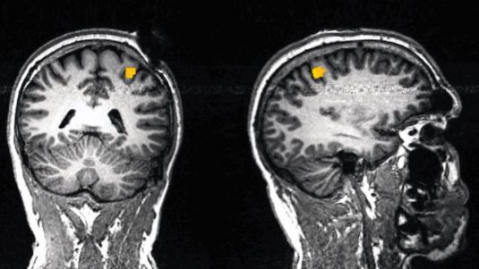
Functional magentic resonance tomography (MRT) shows where ultrasound presumably induces neuromodalation.
As graduate student William „Jamie“ Tyler observed nerve cells under the microscope in the early 2000s, he turned up the bass on his music system. To his surprise, the nerve cells were activated by the sound. Tyler had stumbled on something that had already been reported in the 1950s, but almost forgotten: nerve cells respond to sound. Dr. Tyler, now a professor at Virginia Tech University in Roanoke, (USA) was fascinated by this observation.
Highly invasive procedure
In the meantime, the new technique of optogenetics has revolutionised brain research. For nine years, researchers have been able to selectively activate or inhibit nerve cells in animals. To do this, they alter the genetic material of individual nerve cells in such a way that they can be activated or inhibited by light. This is now an important technique in research on brain function. However, the technique is highly invasive and not yet suitable for application in humans.
„We’ve hit a brick wall with this method“, according to Dr. Tyler. Non-invasive methods for use in humans, such as stimulation of the brain with magnetic fields, are not precise enough. They activate areas of the brain that extend over several square centimetres. „If you aim at the area of the brain that is responsible for movement, you also activate areas that process touch. So specific stimulation is not possible,“ says Tyler. But without specific stimulation, it is impossible to conduct research on the activity of the human brain and its disorders such as Parkinson’s, epilepsy and depression. So scientists are busy searching for a technique that allows them to modulate specific regions of the brain and map it precisely without the use of surgery.
Eight years ago, Dr. Tyler took up the search himself, based on his earlier results. He wrote a proposal and presented it to the patent department of his university: ‚Modulation of brain activity with ultrasound‘. „They said I was crazy,“ he remembers. Tyler looked for the most effective ultrasound frequencies, and eventually tested them in mice. He aimed the device at a tiny area of the brain area that is responsible for paw movement. Nothing happened at first, but then one of his colleagues bumped a switch by accident so that the device transmitted continuously instead of sending pulses. The mouse lifted its paw. „That was the happiest day of my working life,“ said Dr. Tyler. The results were published in 2010, and inspired many researchers to try the technique out.
Millimetre precision
„The low frequencies used by Jamie Tyler produce such a broad pressure field in the brain that it is hard for us as physicists to believe the results,“ according to Jean-François Aubry, of the Langevin Institute in Paris. But Dr. Aubry and his colleagues repeated Tyler’s experiment – with success. They were able to focus the ultrasound stimulus on an area of the brain only a few millimetres in size, and depending on the location of the ultrasound transducer, they could elicit movements of a rat’s whiskers, eyes or tail. Researchers from Stanford and Harvard also reported success. In animal models, they were able to activate the memory centre, inhibit eye reflexes and suppress epileptic seizures.
The ultrasound procedure is similar to that used in the medical diagnosis of patients and pregnant women. An ultrasound probe is placed on the skin and sends brief inaudible pulses into the body. Ultrasound operates at frequencies of over 20 kHz, outside the audible range. The procedure has been in use for over 40 years and is still regarded as quite safe.
It is not clear how ultrasound exerts its effects on the brain. It may alter the structure of the nerve cell membrane. The most abundant components of the cell membrane are phospholipids, and these „rotate or wobble on nanosecond timescales“, as Dr. Tyler notes in a review. Deformation of the membrane by pressure waves changes their arrangement, and channels embedded in the membrane expand like holes in a stretched stocking. This allows charged particles (ions) to flow through the channels, activating the nerve cell. Ultrasound has now been shown to modulate the activity of neurones in bullfrogs, salamanders, rats, rabbits, cats, mini pigs and monkeys.
Researchers could hardly wait to try it out on human subjects, especially because Alexander Bystritsky, a psychiatrist at the University of California, and others have since shown that ultrasound can be combined with imaging techniques such as functional magnetic resonance imaging . So it should now be possible to correlate the type of stimulation with the activity pattern it produces in the brain, and generate a map of brain functions.
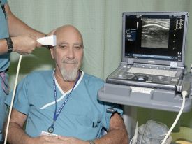
Stuart Hameroff first tried out the effect of ultrasound on himself.
Stuart Hameroff, an anaesthesiologist at the University of Arizona whose research into consciousness is somewhat controversial, was the first to take this step. He started by taking an ordinary ultrasound device, just like the one used in his clinic for diagnosis, and testing it on his own head. Nothing happened. Dr. Hameroff was disappointed at first – but after a minute, he did feel something, as if he had just had a martini. He definitely felt more energised and creative.
In 2013, Hameroff published an initial pilot study in the journal „Brain Stimulation“. He applied 15 seconds of ultrasound to part of the forebrain in 31 patients with chronic pain. Compared with placebo treatment, the mood of patients treated with ultrasound improved and their pain was slightly relieved, although the difference was not statistically significant. Hameroff has since carried out a further study with over 100 participants. In this unpublished study he reports a significant improvement in mood with ultrasound treatment.
Appointments booked up
Neuromodulation with ultrasound is opening up new possibilities. Dr. Aubry plans to find out whether ultrasound can temporarily disable the part of the brain that triggers tremors in patients with Parkinson’s disease. In February, Dr. Bystritsky started a study to see if ultrasound can help people to recover from depression. Dr. Tyler is mapping the brain in animals and humans by combining neuromodulation with imaging. Together with a company he founded, he is also developing a device that should make ultrasonic neuromodulation more practical in patients.
Meanwhile, word has spread that ultrasound may be able to improve one’s mood, and appointments for volunteer subjects in the company’s Boston office are booked for weeks ahead. The next few years will show whether the technique lives up to its promises: a better understanding of human brain function and alleviation of neurological disorders.
printed in: Neue Zürcher Zeitung (January 2014)